AUTHOR: John O’Brien, Chief Revenue Officer
THE PROJECT RISK
A common and costly assumption in an imaging project is that if the scanner fits, the site works.
Medical imaging plays an important role in diagnosis, treatment planning and ongoing care. As access to advanced imaging expands and providers broaden the modalities they offer, the planning requirements of the facility become more complex.
Access to Medicare-funded MRI services is expanding through staged changes to equipment eligibility. Facilities traditionally planned around X-ray and ultrasound may also incorporate CT and MRI and, in larger comprehensive facilities, PET/CT.
Bringing several modalities together creates multiple patient pathways and technical environments under one roof. Each modality has its own equipment, infrastructure, safety and operational requirements, and these need to work as a coordinated service.
A floor plan may show enough space for the equipment. The tenancy may have convenient access, parking and an appropriate local referral market. On an initial inspection, it can appear to be a strong site.
Once the equipment and operational requirements are tested, constraints can emerge. The floor may not support the equipment or shielding loads. The delivery route may be too small. The available power or cooling may be insufficient. A lift, car park, plant room or source of vibration may affect the proposed MRI location. The scanning rooms may fit, but the patient preparation, uptake, recovery, control and equipment spaces may not.
By that stage, the lease may already be signed and the design may be well advanced. What initially looked like a straightforward fit-out becomes a redesign, a negotiation with the landlord or, in the worst case, a site that cannot support the intended service.
A building has to support the service, not just the equipment
A medical imaging facility is more than a collection of scanner rooms. It has to support patients, clinicians, technical staff, equipment, information, supplies and highly specialised building services. The building and facility plan need to accommodate all of these requirements.
This becomes more important as facilities add modalities. A facility combining general X-ray, ultrasound, CT, MRI and PET/CT needs to accommodate several different patient journeys and technical environments. Some patients will move quickly from reception to an examination room. Others may need to change, drink contrast, be cannulated, undergo MRI screening, spend time in a PET uptake room or be observed after a procedure. Treating every modality as a room with a machine inside it misses much of what determines whether the facility will operate successfully.
The building requirements that are easy to underestimate
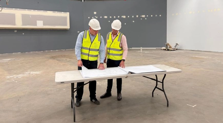
Several building requirements can materially affect site suitability, capital cost and the delivery program, even when the equipment appears to fit on an initial plan. They need to be considered alongside the equipment layout, not after the design has been fixed.
Equipment access and replacement. The delivery path needs to be tested from the site boundary to the final equipment position. Loading areas, door openings, corridors, lifts, corners and temporary wall removals must work for the initial installation and eventual replacement. It is not enough to find a way to install the equipment once if removing it later requires major disruption to the building or an operating facility.
Structure and buildability. Floor loading, slab condition, ceiling height, penetrations and structural supports need to accommodate the selected equipment. Shielding and shielded doors can add significant loads. Ceiling-mounted X-ray equipment, injectors and services may also require support that is not present in a standard commercial tenancy.
Power, cooling and technology. Imaging equipment and chillers can place significant demands on electrical and mechanical infrastructure. Computer cabinets, workstations and reporting systems introduce additional power, cooling, data and connectivity requirements. Electrical capacity, redundancy, heat rejection, plant space and cable routes should be tested against the selected equipment and operating hours. RIS/PACS integration also needs to be confirmed.
Safety and shielding. Radiation shielding for CT, PET/CT and X-ray must respond to the selected equipment, anticipated workload and the use of surrounding areas, including spaces above and below. MRI requires a controlled magnetic environment, radiofrequency shielding and assessment of the magnetic fringe field. These are site-specific design inputs, not standard details that can simply be transferred from another facility.
The space outside the scanning room. Reception, waiting, changing, screening, patient preparation, uptake, holding or recovery, toilets, storage, utilities, staff work areas and reporting all contribute to the required footprint. A plan that protects the scanner room by squeezing these areas usually transfers the constraint into patient flow and staff workflow.
Different modalities expose different site risks
Early planning does not need to resolve every technical detail. It should identify anything that could materially affect whether the site is viable.
MRI. The assessment extends beyond the magnet’s size and weight. It needs to consider floor capacity and levelness, installation and replacement access, vibration, external electromagnetic interference, the fringe field, controlled access, RF shielding, power, cooling and the equipment-specific emergency provisions. Nearby lifts, moving vehicles, plant and sensitive equipment can influence where an MRI can be located even when the room dimensions appear suitable.
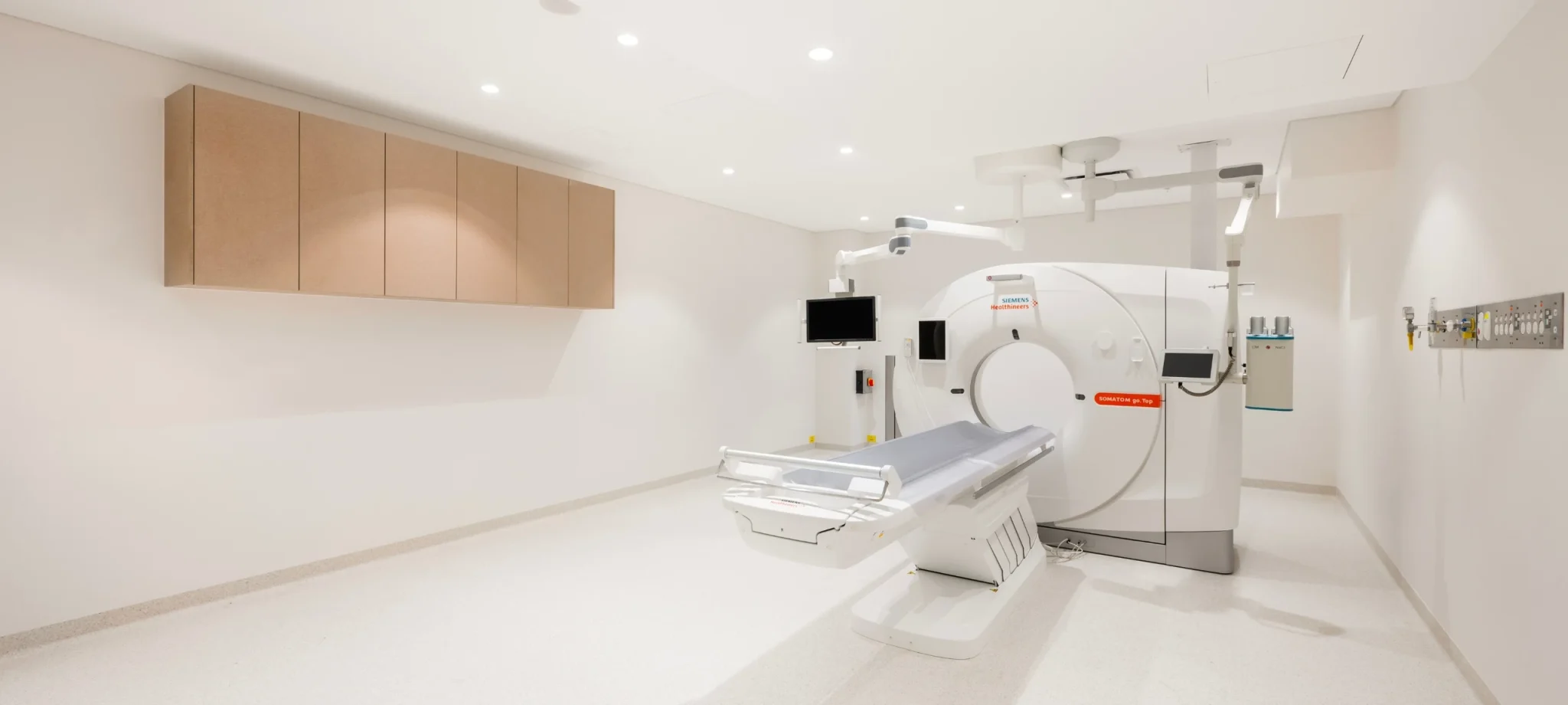
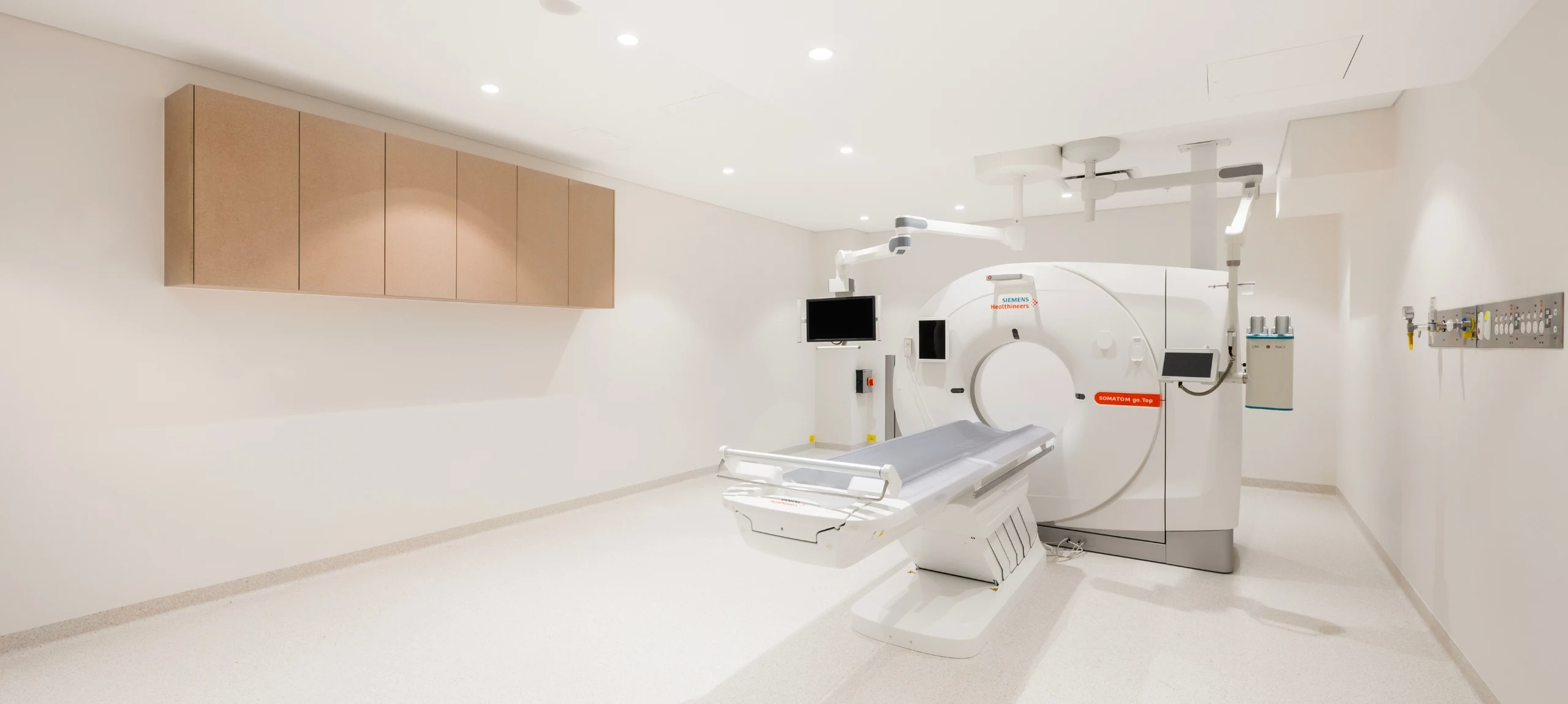
CT and X-ray. Both use ionising radiation and require shielding designed for the selected equipment, anticipated workload and surrounding occupancy. X-ray shielding must also account for beam direction. CT also brings substantial power, cooling, structural and equipment-room requirements. The clinical model may add contrast preparation, cannulation, accessible changing, toilets, observation or recovery. X-ray rooms may require structural support for ceiling rails and wall-mounted equipment as well as suitable patient and operator circulation.
PET/CT. Where PET/CT is included, it is not simply a CT scanner with additional shielding. The use of radiopharmaceuticals can require uptake rooms, controlled patient holding, separation of dosed and undosed patients, a dedicated toilet, radiopharmaceutical receipt or preparation, waste storage and carefully managed staff and patient movement. The number of uptake rooms and the time patients spend within the service influence throughput and can make the overall footprint materially larger than a conventional CT suite.
Ultrasound. Ultrasound does not require radiation shielding, but the room still needs to support sonographer ergonomics, examination-bed access, lighting control, privacy, handwashing, power and data. Access to patient toilets is important for full-bladder examinations, while procedures may introduce additional space, storage, recovery and transducer reprocessing requirements. Poor ultrasound planning may be less visible during construction, but it is felt by staff every day after opening.
There is no single answer to how much space is needed
Healthcare planning guidelines and vendor information provide useful benchmarks, but neither can determine the footprint without understanding the service. The same scanner can be installed within very different facilities depending on patient volumes, case mix, procedures, staffing and whether the facility operates as a standalone modality or a comprehensive service.
Generic room schedules are useful, but they are only a starting point. The final area needs to account for the equipment and its clearances, control and equipment rooms, patient and staff support, servicing, circulation and the ability to replace equipment or introduce additional services later. Designing to the smallest possible footprint can reduce the initial tenancy area while limiting capacity, privacy, efficiency and future flexibility for the life of the facility.
What should be confirmed before committing to a site?
Before committing to a site, the operator and project team should understand the questions capable of changing the decision:
- Clinical fit. Can the site support the proposed modality mix, patient journey, staffing model and expected activity?
- Planning and approvals. Do the permitted use, operating conditions and likely approval pathway support the proposed service?
- Equipment and structure. Can the building accommodate the selected equipment, shielding, structural loads and ceiling requirements?
- Delivery and replacement. Is there a workable route to install, service and eventually replace each major item of equipment?
- Building services. Are the available power, cooling, plant space, data and other services sufficient, or can they be upgraded without unacceptable cost or program impact?
- Safety and neighbouring uses. Can radiation shielding, MRI controlled areas and other safety requirements be achieved without creating unacceptable impacts elsewhere in the building?
- Future flexibility. Can the facility replace equipment, add capacity or introduce new services as demand and technology change, or will the site reach its physical limit soon after opening?
The value is in coordinating the decisions
The individual requirements of medical imaging are generally well understood within their disciplines. Equipment vendors provide technical planning information. Healthcare planning guidelines describe planning principles. Radiation experts, engineers and regulators establish the relevant safety and compliance requirements.
The difficulty is that the requirements are interdependent. Changing the equipment can alter power, cooling, shielding, structure and room geometry. Moving the scanner can affect delivery, workflow, services and approvals. Adding PET/CT can change patient circulation and the support-space calculation for the wider facility.
A technically valid decision made by one discipline can create an operational or commercial problem somewhere else.
Coordinated planning identifies constraints early and tests their combined effect on site suitability, cost, program and future operations, reducing the risk of redesign, variations, delays and unexpected costs.
PUTTING IT INTO PRACTICE
The right question is not simply whether an MRI, CT, PET/CT, X-ray or ultrasound unit will fit inside a tenancy. It is whether the building can support the complete imaging service: the equipment, patient journey, staff workflow, infrastructure, compliance requirements and future direction of the facility.
Testing the clinical model, equipment and building together before commitments are made identifies risks to safety, operational efficiency, cost, program and future flexibility while the project still has options.
Perfect Practice brings site assessment and project due diligence together with facility planning, design and delivery. This helps clients test site suitability, identify constraints early and establish a clearer path from site selection to opening.
 AUTHOR: JOHN O’BRIEN, Chief Revenue Officer
John has more than 22 years of clinical, operational and commercial experience in healthcare. Clinically trained as a nuclear medicine scientist, he has held senior operational and executive leadership roles within a national medical imaging group. His experience spans clinic operations, new site developments, refurbishments, service expansions and the integration of acquired businesses across a national network.
As Chief Revenue Officer at Perfect Practice, John leads the company’s revenue strategy across business development, client relationships and marketing, with a focus on sustainable growth and long-term client partnerships. He works closely with clients and internal teams from early engagement and project planning through to delivery, helping ensure commercial objectives, operational requirements and project outcomes remain aligned.
John holds qualifications in Nuclear Medicine and an MBA.
Connect with John on LinkedIn